

How To Clean Out A Scab Stuck In Superior Turbinates
![]() Open admission peer-reviewed chapter
Open admission peer-reviewed chapter
Management of Common Complications in Rhinoplasty and Medical Rhinoplasty
Submitted: August 18th, 2022 Reviewed: March 16th, 2022 Published: August 31st, 2022
DOI: 10.5772/63130

IntechOpen Downloads
2,691
Total Chapter Downloads on intechopen.com
Abstruse
Rhinoplasty is considered among the nigh challenging aesthetic operations because many variables accept to exist taken into consideration to achieve an optimal aesthetic and functional result. This implies that complications are always waiting effectually the corner. Information technology is of prime importance to know the chief minor and major complications related to the process to be able to prevent and care for them promptly when required. Septorhinoplasty is a delicate and difficult procedure, which requires authentic anatomical cognition and important clinical experience. Withal, complications can affect both inexperienced and expert surgeons. Thus, the most frequent complications of rhinoplasty should exist known and adequately prevented when possible.
Keywords
- Adverse events
- rhinoplasty
- complications
- rhinofiller
- Medical Rhinoplasty
*Address all correspondence to: storres100@gmail.com
1. Introduction
Some mail-operative complications are easily treated, whereas others require multiple reconstructive surgeries and sometimes
Advertisement
2. Traumatic complications
two.1. L-construction or 1000-area fracture
During septorhinoplasty, whatever approach is used, ii cardinal rules must exist kept in mind:
-
Respect the Cottle K expanse, which is anatomically divers as the intersection of the nasal bones, septum and triangular cartilages.
-
Preserve an adequate dorsal-caudal L structure for support.
Damage to these structures causes an inadequate support of the nasal pyramid and with time causes nasal dorsum collapse and back sill deformity spontaneously or after modest trauma. An adequate dorsal-caudal L structure of at least 1 cm is necessary for structural support to prevent this blazon of complexity. The K area should be addressed with extreme care upon dorsal hump removal. Precise subperiosteal dissection is done above the nasal bones with a Joseph dissector. Incremental dorsal hump reduction with a rasp or osteotomes allows for maneuver control and removes the difficult tissue while avoiding impairment to the triangular cartilages or nasal bones.
2.2. Dental trauma
2.iii. Intracranial complications
Intracranial complications include rhino-liquoral fistulas and anosmia.
Given the fact that the superior portion of the septal cartilage is direct abutting the cribriform lamina of the ethmoid and is the direct continuation of this structure, an understanding of why this severe complication is not that uncommon is apparent.
Septoplasty is a fragile phase of the procedure. Very oft, surgeons care for the septum aggressively by grabbing the bony portion with Weil forceps and attempting to break or remove the tissue through rotatory movements. Prevention of rhino-liquoral fistulas consists of an accurate and delicate septum autopsy, peculiarly with regard to the superior bone portion. Before pulling a fragment, adequate dissection and freeing is necessary. The clinical symptomatology of rhino-liquoral fistulas includes rhinorrhea and positional cephalus. Diagnosis is confirmed through beta-2 transferrin presence in the fluid, specifically the cerebrospinal fluid.
Such complexity requires hospitalization, lumbar drainage positioning by a neurosurgeon and eventual multilayer nasal endoscopic fistula repair.
Anosmia is fortunately very seldom observed due to the damage of the olfactory bulb. Near frequently, this condition is secondary to a persistent respiratory nasal obstructive pathology.
2.4. Orbital complications
Orbital complications related to septorhinoplasty are extremely rare and include blindness and epiphora. Blindness has been reported in some cases and is related to turbinate or nasal dorsum steroid injections. The etiopathogenesis described involves an embolic occlusion of the central retinal avenue. Other cases due to vasoconstrictor injections in the septum and turbinates, being the etiology of a spastic response on the key retinal artery, have been described [1–3].
These unfortunate complications are hard to predict and impossible to resolve. For this reason, prevention is washed by fugitive steroid infiltration in the turbinates and aspiration prior to injection and injecting a small quantity when treating the dorsum. Epiphora is an extremely rare complication after rhinoplasty. Lateral osteotomies are generally safety if executed in a standard manner. Impairment to the lacrimal ducts is possible when the osteotomy direction is incorrect or when motorized instruments or saws are used. Information technology is frequently clinically confused with paralateronasal edema. Spontaneous resolution is oftentimes verified, although desultory cases crave dacryocystorhinostomy for complete resolution [4, five].
Advertisement
iii. Respiratory complications
3.1. Internal nasal valve dysfunction
Internal nasal valve dysfunction is a frequent complication secondary to sometime schoolhouse destructive rhinoplasty. The principal cause is over-resection of the lateral cartilages during hump removal. The internal nasal valve bending is formed by the confluence of the nasal septum medially and lateral cartilages externally; its normal value is around 15° [6].
A reduction in this value determines damage in airway menstruation. More than severe than an excessive resection of the triangular cartilages is scarring in the internal valve area due to transmucosal disjunction of the septum from the triangular cartilages; fortunately, this is an old and discarded technique. Patients with a not-deviated septum are referred for treatment of severe nasal respiratory problems. Moreover, in addition to this severe functional defect, a dorsal inverted Five deformity appears after the resolution of the surgical edema due to inferomedial plummet of the triangular cartilages [seven].
The remedy for this blazon of complication is the placement of a spreader graft, whatever the type (motorcar, mini or classic) and source (septum, concha or rib). The of import technical item is to place the graft so as to raise and reposition the collapsed triangular cartilages and return internal nasal valve function, augmenting the cantankerous-sectional area.
Spreader grafts also allow straightening of a cephalic deviated septum, reconstruction of an open roof deformity or improvement of dorsal artful lines [8].
three.2. Nasal septal perforation
The etiology of septal perforation is various and may be iatrogenic, which is nigh oftentimes the instance, due to cocaine abuse, infections, trauma and granulomatous diseases. With subperichondrial septum dissection, circumspection should exist taken not to trespass the mucoperichondrial flaps. Information technology is advisable to start with the easier side to grant integrity in at least i side. When both mucosal flaps are damaged bilaterally, an iatrogenic septal perforation will exist produced.
The symptomatology of septal perforation includes crusts, recurrent bleeding, whistling or inspiratory rumors and nasal respiratory obstruction. The more than anterior the perforation is, the greater the associated disturbance.
The virtually ancient solution for the trouble was the utilize of silicone septal buttons, which are less popular amid patients nowadays.
Diverse septal perforation repair techniques have been described, with the most effective ones being from Kridel and Castelnuovo [ten, 11]. Kridel described an open approach for the provision of sliding superior (from the internal nasal valve region) and inferior (from the nasal flooring and inferior turbinate) mucoperichondrial flaps. Castelnuovo reported an endoscopic arroyo for an intranasal septal mucosal pedunculated flap to the ethmoidal arteries, which is rotated to obtain defect closure. Both techniques grant a high success charge per unit.
Whatever the case, it is proper to preclude septal perforation and if verified to take time to repair the mucoperichondrial flaps properly. Allotting an additional 10 minutes at the main surgery is better than performing three hours of revision surgery for perforation closure.
3.three. External nasal valve dysfunction
The external nasal valve is an area defined iii-dimensionally by the inferior turbinate head, caudal portion of the triangular cartilages, cephalic portion of the alar cartilages and septum. The most common source of post-rhinoplasty dysfunction is related to an excessive resection of lateral crura of the alar cartilages.
This condition is occasionally seen when an attempt to reduce nose tip dimensions is sought at all costs, not leaving enough alar cartilage to support the nasal ala.
Nasal alar collapse tin exist dynamic if information technology manifests just during inspiration (forced or not) or static in more than severe cases if it is verified at residual. The minimum alar cartilage dimension to preserve varies according to the intrinsic consistency of the cartilage and it is non the same for all patients. Nevertheless, a minimum of iv–5 mm should exist kept and old risky, interruptive approaches should exist avoided.
Multiple techniques have been described to treat this complexity, namely, alar spreader grafts, lateral crura repositioning, alar spanning grafts, barrel whorl technique, lateral crura strut grafts and alar batten grafts. Alar batten grafts are the nigh frequently used, but every case should be analyzed individually and treated appropriately with the nearly indicated technique [12].
External nasal valve compromise is also verified after maneuvers that cause narinal stenosis. This condition is seen, for example, when a sloppy adaptation of the vestibular pare occurs later rhinoplasty due to a lack of closing sutures in the surface area, infection or abnormal scarring. Another cause is represented past excessive alar base wedge resection.
Corrections in these cases are complex and foresee the use of local flaps and Z-plasties, simply in the majority of cases, auricular composite grafts are necessary to replenish the lack of previously excised tissue.
Residual anterior septal deviations and turbinate hypertrophy can cause external nasal valve dysfunction. Residual anterior septal deviations require surgical revision with a more precise septoplasty. Inferior turbinate hypertrophy is very frequent, especially in allergic patients. In these cases, medical therapy is advised with local steroids and systemic antihistamines, discouraging continuous surgical retouching [13].
Turbinoseptal synechiae (adherences) can also produce external nasal valve stenosis, although they can appear even more posteriorly in the nasal fossae. Silicone splints should be used and kept in identify long enough to allow re-epithelization of the turbinate and septum to prevent turbinoseptal synechiae when mucosal lacerations occur.
iii.4. Sinusitis
Sinusitis is rare equally a post-rhinoplasty complication, only it can become credible if unrecognized predisposing conditions are nowadays.
The medial meatus protected by the middle turbinate represents the common drainage path for the ducts of paranasal sinuses. Ethmoidal anterior, frontal and maxillary sinuses all drain at this level.
Medial turbinate lateralization maneuvers are extremely dangerous as they may cause rhino-liquoral fistulas and compromise normal paranasal sinus part.
If predisposing conditions are present, the presence of concha bullosa may predispose a patient to post-rhinoplasty sinusitis. Information technology is advisable to appraise pre-operative nasal and paranasal sinus CT scans that will give valuable information regarding septal deviation and turbinate hypertrophy and identify sinusal alterations suitable to be treated during the surgery through functional endoscopic sinus surgery (FESS) to avoid this complication.
Advertisement
4. Aesthetic complications
4.i. Supratip deformity (polly bill)
Post-operative deformity of the supratip nasal surface area that assumes a convex shape in relation to the nasal dorsum can have ii sources: cartilaginous tissue or scar tissue. Cartilaginous polly beaks are caused by an insufficient resection of the inferior third of the dorsal septum in proximity to the septal angle. Scar tissue polly beaks, on the other hand, are more than frequent in cases with sebaceous skin and are due to hypertrophic scarring of the subcutaneous tissue of the supratip region.
The remedy for supratip scarring is based on local steroid injections; they are very effective if done properly with regard to timing and modality. Triamcinolone acetonide (Kenacort, xl mg/ml injectable suspension) is the steroid of option. Dosage should be triamcinolone i–2 mg practical early (ii–three weeks afterward the surgery) if a tendency for supratip deformity is perceived and non repeated before a ii-month interval. The effect of the therapy is seen in the following 2 months mail injection. The injections should be in a deep plane and never intradermal. Superficial injection causes cutaneous atrophy, telangiectasia, depressions, color modifications and underlying cartilage visibility [15].
Cartilaginous supratips and not-responders with scar-based supratips are treated with revision surgery. An in-depth analysis of tip-dorsum relation and the use of tip-defining grafts (onlays and shields) are useful to avert recidivism.
4.2. Dorsal irregularities
The nasal dorsum is the region more than decumbent to unexpected and unwanted surprises after a rhinoplasty. It is very hard for the surgeon to ensure that no dorsum unevenness remains at the end of surgery and that the end result is smooth and with no imperfections in the majority. Nevertheless, months or years afterwards the surgery, information technology is difficult to observe an operated nose that does not show some dorsal irregularities at least upon palpation. The reason for this is that surgical edema will hide modest irregularities and mask an acceptable palpation evaluation of dorsum smoothness. With fourth dimension, as nasal tissue swelling disappears, irregularities outset to show [16].
Dorsal deformities are among the most mutual causes of revision rhinoplasty. They are generally due to excessive or inadequate hump removal, remnant fragments later removal, asymmetric resections, inadequate graft modeling or fixation and dislocation.
Open up rhinoplasty can reduce the frequency of these imperfections as information technology allows for straight vision of the back. Another tip to reduce the percentage of these complications is to perform dorsal index palpation with the surgical gloves wetted with normal saline, augmenting sensitivity for the surgeon. Profuse cleansing and washing of the dorsal area under the skin envelope before suturing is imperative as it eliminates pocket-sized cartilage residues and bony fragments, avoiding future irregularities.
Fugitive dorsal irregularities in patients with thin skin is notwithstanding very difficult. In these cases, it may be advisable to utilise dorsal augmenting materials. These can be autologous (temporal fascia, perichondrium graft), heterologous (equine or bovine pericardium membranes) or alloplastic (Gore-Tex). Autologous materials are preferable due to the lower incidence of infections associated with them; still, at the back level, the take chances of infection or extrusion is very small even for not-autologous materials [17].
4.3. Tip deviations and irregularities
Another particularly anti-aesthetic condition is an altered tip projection, either hyperprojection or hypoprojection. Nose tip deformities often manifest a long time afterwards the surgery (1 or 2 years after). In fact, the nose tip is the concluding region to swell downwards in the mail service-operative flow.
Prevention of this complication relies on noesis of tip supportive mechanics and the tripod theory too as attention to avoid confusing or destructive techniques. Nevertheless, the well-nigh important factors are still expertise and respecting artful proportions that will grant expert results in the long term. Revision rhinoplasty is surely easier and anticipated if done via an open up approach, but this also depends on the skills and experience of the surgeon [20].
iv.four. Pare necrosis
Nasal pare necrosis is among the worst complications that can occur during a septorhinoplasty. It is mainly acquired past vascular damage in the vessels that supply the nose tip. Rarely, information technology tin can nowadays due to excessive dressing compression. Most frequently, it appears later on damage in the lateral nasal arteries due to an incorrect plane of autopsy or post-obit excessive olfactory organ tip fat tissue reduction, in an try to reduce its size.
A new source of peel necrosis of increasing prevalence is the post-operative use of dermal fillers at the nasal pyramid, nasolabial folds or paranasal region to camouflage irregularities. This issue is more frequently verified when the filler is delivered with needles that may cause direct vessel harm and intravascular occlusion or indirect vascular compression, jeopardizing tip vascularity.
Some rules should be respected to prevent this complication:
-
Avoid injecting fillers with sharp needles (preferably blunt tip cannulas) in paranasal areas.
-
Dissect the nasal tissues attached to the cartilaginous framework without getting superficial.
-
Avert defatting techniques of the nose tip or reduce it to a minimum.
-
Avoid firm and tight dressings, particularly in revision cases.
-
Limit alar wedge resections under the alar crease.
Treatment of skin necrosis is very complex and ranges from bourgeois approaches (such as 2nd-intention wound healing) to complex reconstruction procedures with local, regional or gratuitous flaps. Whatsoever the approach, skin tropism and elasticity are a primary goal before intending more complex repair. The latter process tin exist accomplished through platelet-rich plasma and micro-lipofilling sessions.
Advertisement
5. Infective complications
Rhinoplasty infections are non frequent, probably due to the natural protective mechanisms of the nasal mucosa. Even so, the myriad of infective cases tin exist very vast and get from small subcutaneous cellulitis due to infected sutures to severe cavernous sinus thrombosis.
Local pare or mucosal infections are treated with local and systemic antibiotics. Abscesses may affect the dorsum, tip or septum, with septal abscess beingness the well-nigh unsafe, and they should be promptly tuckered; septal abscess can appear from an undiagnosed septal hematoma that can evolve to a septal perforation if non treated promptly.
High fever, meningeal signs, nausea, vomiting and hypotension are suggestive signs of a severe infection, such equally cavernous sinus thrombosis. If the diagnosis is suspected, nasal tampons should be removed immediately (especially if placed several days before) and secretions should be sent for cultural and bacteriological analysis, with the about frequent germ involved being
Safety antibiotics in rhinoplasty are a controversial topic but still highly indicated past the majority of surgeons.
Advertisement
6. Vascular complications
Vascular complications include septal hematoma and epistaxis.
vi.ane. Septal hematoma
Septal hematoma can occur secondary to trauma or surgery and is a serious complication. Its symptomatology includes nasal obstruction, pain and, occasionally, fever. Inductive rhinoscopy reveals a septal mass that occludes one or both nasal fossae. Immediate therapy is indicated and consists of hematoma drainage, nasal tampons to impede recidivism and proper antibiotic therapy to avoid abscess transformation.
Septal abscesses tin can evolve to mucosal and/or cartilage necrosis and septal perforation varying in dimension and location according to the underlying infection [21].
6.2. Epistaxis
Bleeding in rhinoplasty patients post-operatively is normal if limited, whereas it can become a complication if profuse or continuous. The status is more frequent in at-chance patients on anticoagulants or platelet anti-aggregating agents. In these cases, prior consultation with a hematologist and a cardiologist is appropriate, and oral clot-altering drugs should be discontinued and subcutaneous LMW heparin initiated several days before the surgery. All patients should exist brash to discontinue NSAID or aspirin intake at least 2 weeks prior to functioning.
A precise and delicate technique during surgery is desirable to avoid vascular problems. During septoplasty, for example, it is of import to avoid mucosal flap lacerations to minimize bleeding. A nasal septum mattress suture can exist useful to foreclose bleeding and septal hematoma. Turbinate cautery should be gentle. An open technique allows for direct vision and hemostasis of bleeding vessels during the procedure. Epistaxis therapy includes 60° head elevation, nasal packing and gentle nares pressure for x–15 minutes. Severe epistaxis tin crave an emergency endoscopic process to coagulate the sphenopalatine septal and lateral branches.
Advert
seven. Medical rhinoplasty
Medical rhinoplasty was first described past Braccini and Dohan Ehrenfest [22] in 2008. The concept, although highly polemical and refused by rhinoplasty surgeons at its onset, developed popularity among artful patients due to its minimally invasive characteristics, with minimal or no downtime and pleasing aesthetic improvements.
The term
vii.1. Rhinofiller
Rhinofiller specifically involves the infiltration of a dermal filler to modify external or internal nasal structures for aesthetic or functional purposes. Since its introduction in 2008, many temporary and permanent substances have been used to accomplish the desired corrections. Successful application mandates acceptable anatomical knowledge of the related structures.
Proper patient selection is of import to achieve good results. Exclusion criteria include severe nasal airway impairment, permanent filler in the area, history of ischemic/thrombotic events or known hypercoagulability, local infection and recent trauma.
Before the procedure, nasal assay should be performed clinically and photographically to define needed corrections.
Areas of potential correction include dorsal artful lines, the back, minor hump camouflage, radix enhancement, tip rotation and projection and base of operations augmentation. Details are shown in Effigy 11.
Functionally, in selected cases, the use of fillers can be useful to augment the aperture of the internal nasal valve as a volumetric spreader graft.
Morphing simulations are advisable before treatment in gild to give patients an indication of the post-treatment outcomes, explain the process and constitute common goals. In addition, specific, informed consent should be properly discussed and obtained.
7.1.1. Technique
Treatments are typically performed with medium-viscosity hyaluronic acid (HA) fillers nether local coldhearted (lidocaine intradermal vesicles applied using a 0.three ml syringe with a 32G needle) with the aid of a 25G (0.5 mm) × 4 cm blunt-tip dispensable cannula, manually bent, maintaining sterility at all times, in order to obtain improve compliance of the shapes and silhouette inside the nasal area. The distribution of fabric should be performed as required to follow the treatment programme. Tip refinements can exist sporadically carried out through needle infiltration with extreme care.
The specific design of anesthetic peripheral blocks and filler infiltration is shown in Figure 12.
Generally, the patient satisfaction rate with this correction is very high and, due to the scarce muscular action in the nose, corrections with Hyaluronic acid dermal fillers terminal more than 1 twelvemonth and in many cases even 2 years. A clinical example of rhinofiller is described in Figures 13 and 14.
Advertisement
eight. Discussion
The nasal area is composed of different interacting tissues, such as the peel, subcutaneous tissue, muscle, bone, cartilage and mucosa, which come together to course a normal, functional and aesthetically pleasing nose. To make things more complicated, in that location is as well a vascular anatomy formed by two master circuits, namely, the supratrochlear and dorsal arteries and the facial circuit that includes the superior labial and angular arteries, all of which are anastomosed in the tip. This has been the field of study of recent involvement and written report because information technology is believed that a proper technique and anatomical knowledge are of prime importance in order to avoid vascular complications [28–30]. Facial vascular complications were start described in 1991 after collagen injections in the glabellar area [31]. The reported incidence of Nicolau syndrome or embolia cutis medicamentosa (ECM) post-obit glabellar treatments is 9/10,000 procedures (0.09%). The known risk factors associated with this catastrophic issue are a high syringe piston pressure, a highly vascularized territory and previously traumatized tissue. The outset of these factors tin be mitigated using fluid materials of depression viscosity. Unfortunately, the entire facial region, particularly the nasal area, is considered highly vascularized and many reports of paranasal vascular complications, which vary from mild symptoms of pain and skin color changes to necrosis and even bilateral blindness, have been published [32–41]. The pathophysiology of ECM is an intravascular injection that advances in a retrograde mode to a distant area and, through changes in blood pressure, arrives at a distant vessel and causes a vascular complication. The resulting symptoms vary according to the physiology of the vessel that is compromised; affliction of arteries leads to pallor, whereas apoplexy of veins manifests equally livedo reticularis. According to the author's feel, there is a second mechanism of vascular compromise in the nose known as
-
Patient selection: Proper patient selection is vital in society to achieve a skilful consequence. Rule out individuals with unrealistic expectations and treat mail-rhinoplasty patients with extreme care. -
Materials: A good technique begins with selection of the correct materials. Only temporary or autologous materials (fat) should exist used in the olfactory organ. Among temporary materials, HA is the best option because it causes no fibrotic changes in the subcutaneous tissue, such as those that tin occur with calcium hydroxyapatite. Moderate-viscosity HA is preferred due to the lower piston force per unit area in the syringe associated with it. -
Correct amount of textile: Never exceed the correct quantity of filler used in the nose. Information technology is always meliorate to undercorrect and then echo equally needed. A good rubber measure is to stay within one ml of filler per session. Recollect that the pressure of the material tin can induce vascular problems even without being intravascular. Identify the fingers to position and maintain the product in the target area to avoid migration. Small amounts of material should be placed using depression infiltrative pressure level and few passes in a retrograde infiltration fashion. -
Cryotherapy: It is ever wise to favor vasoconstriction in social club to limit bruising and edema and reduce intravascular compromise. -
Cannula, manually curved: The utilise of atraumatic cannulas permits gentle dissection of the tissues, reduces the trauma and risks of intravascular injection and delivers the material through a laminar flux that guarantees evenness. The manually curved feature allows for perfect shape compatibility with the nasal dorsum. The use of local anesthetic vesicles and needle skin penetration prior to cannula entry limits pain, trauma and vascular compromise. -
Needles: Extreme circumspection should be used when injecting with needles effectually the olfactory organ; their use should be express to retouches or refinements and but by very experienced physicians. Perform tunnels (visible entry and exit points created with the needle being used) and allow material to exit if needed. The virtually risky areas are the tip, glabella, canine fossa and columellar base. Avoid bolus techniques in these regions and inject only whencoming out . Information technology is preferable to use medium-sized needles and inject into the deep or intermediate airplane. Prior aspiration is non useful. -
Improve; practise non effort a perfect outcome: This technique should exist considered part of the armamentarium of every aesthetic surgeon just not used as a single instrument. Whenever we want to completely correct a surgical deformity with fillers, nosotros get into possible complications. -
Planning and discussion of potential complications: It is essential to obtain proper informed consent. Oft, patients are ill-informed about this procedure and have often read that it is extremely easy and free of risks. Establish a good human relationship based on truth and trust with your patient. Morphing software tin can be of peachy help in this phase to help communicate with patients and establish common goals; underpromise and overdeliver. -
Analyze the columellar labial angle: Analysis of this feature allows for objectivity of the outcome and fifty-fifty the most disquisitional patients will be able to appreciate the improvement. -
Available kit for potential ECM: If you intend to care for the olfactory organ with dermal fillers, you should exist prepared to handle the complications as well.
Advertising
9. Conclusions
The employ of dermal fillers effectually the nose, although an avant-garde technique with potentially severe adverse events, is a powerful tool that can be used with a keen deal of satisfaction and prophylactic for the do good of patients who wish to accomplish aesthetic or functional improvements without a surgical procedure. The risks and benefits should always be considered and discussed, and complications should exist prevented and promptly treated if necessary.
9.1. Nasal botulinum toxin
The onset of the neurotoxin in aesthetics revolutionized the handling of dynamic facial dynamic wrinkles, producing a reversible paralysis that allows overlying tissues to relax and aesthetically to be flattened and raised. The use of botulinum toxin around the nose differs from the typically recommended indications of the superior facial 3rd, being considered an advanced and off-characterization technique.
The use of botulinum toxin in the olfactory organ is useful in hypermotile noses that typically motility with mimic expression. The complications related to this technique are non as severe equally those associated with the use of rhinofiller as they are reversible and practise non affect nose vascularity. Complications include pain, bruising, swelling, asymmetries, brusk-lasting outcome and resistance. The duration of the corrections is express (three–iv months) and activeness takes ii–ten days to establish, but it may raise the results obtained with a rhinofiller as information technology removes muscular action and tension over the nasal region. Deep punctures at a muscular level are necessary.
The following muscles suitable for handling around the nose are as follows:
-
The
nasalis transverse muscle is responsible for the wrinkling in the radix paranasal region known asbunny lines . Treatment typically requires 1–two U per side one mm higher up the angular vessels at the lateral attribute of the radix. -
The
levator anguli oris alaeque nasi muscle is responsible for gummy smiles. Treatment requires 2–v U per side at the intersection of the nasolabial fold and the alar region. -
The
depressor septi nasi muscle is responsible for hypermotile nose tips and an acute columellar labial angle. Treatment requires 1–2 U at the base of the columella. -
The
alar nasalis muscle acts together with the depressor septi nasi muscle to lower the tip projection and restrict the nasal discontinuity. Treatment requires ane–2 U per side at the midpoint of the alar area.
A summary of these muscles and their corresponding treatment doses is given in Figures 15.
Advertisement
10. Clinical instance patients
x.1. Clinical case patient 1
A 28-year-old female patient who previously underwent destructive septorhinoplasty with excessive resection of the alar and triangular cartilages presented to us with an inverted Five deformity, right nasal alar collapse, tip asymmetry and a deformed back sill.
Revision rhinoplasty was done using an open approach and harvesting right concha cartilage grafts. Tip de-project, right lateral reconstruction and bilateral spreader graft positioning were performed (Figures 1–4).
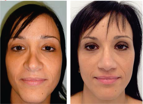
Figure 1.
Clinical case 1: Earlier (correct) and later on (left) images.

Effigy 2.
Clinical example 1: Before (upper) and later (below) images.

Effigy 3.
Clinical case 1: Before (left) and after (right) images, lateral view.

Figure iv.
Clinical case 1: Before (left) and later (right) images, oblique view.
10.2. Clinical instance patient 2
A 42-yr-one-time female patient who previously underwent septorhinoplasty presented to united states of america with dorsal irregularity, tip disproportion and a 3 cm diameter anterior septal perforation.
Reconstructive procedure was performed using an open approach and the Kridel septal perforation closure technique. Regularization of the dorsum and tip symmetrization was done (Figures 5–viii).

Effigy 5.
Clinical example 2: Septal perforation.

Figure 6.
Clinical example 2: Before (right) and after (left) images.

Effigy 7.
Clinical case ii: Before (upper) and after (lower) images, basal view.

Figure 8.
Clinical example two: Before (right) and later on (left) images, lateral view.
10.three. Clinical case patient three
We also report the case of nasal lipofilling for iatrogenic skin necrosis post-rhinoplasty and filler use in a 22-year-old female patient who previously underwent open rhinoplasty and received several steroids and filler (HA) treatments in the post-operative menstruum until the olfactory organ tip, alar cartilages, caudal septum and anterior nasal spine vascularity were jeopardized. The patient was referred with astringent scarring and low skin elasticity. She refused reconstruction with a forehead flap. Our handling plan was initiated with PRP mesotherapy to the nasal region through a dermic pen device. Successive nasal micro-lipofilling sessions (×4) enhanced with a xx% mix of PRP significantly improved skin quality and elasticity for farther reconstructive steps (Figure ix).

Figure nine.
Clinical instance three: Dramatic ischemic progression due to fillers and steroid injections post-rhinoplasty courtesy of Dr Sebastian Torres.

Figure 10.
Clinical case iii: Micro-lipofilling technique (left) and mail service-operative (12 months) reconstructive procedures (centre and right) courtesy of Dr Sebastian Torres.

Figure 11.
Rhinofiller primary treatment areas.

Figure 12.
Rhinofiller injection technique. Spots indicate the entry point for cannula; orange triangles bespeak material distribution.

Figures 13-14.
Rhinofiller pre-operative (upper) and post-operative (lower) firsthand results, lateral view.

Effigy 15.
Summary of paranasal muscles and botulinum toxin doses.
References
- 1.
Rees TD. Aesthetic Plastic Surgery. Philadelphia, PA: WB Saunders; 1980. - 2.
Paun SH, Nolst Trenité G. Revision rhinoplasty: an overview of deformities and techniques. Facial Plast Surg 2008; 24 (3):271–287. - iii.
Adamson PA. The failed rhinoplasty. In: Current Therapy in Otolaringology Head and Neck Surgery. Toronto, ON: BC Decker; 1990: 137–44. - 4.
Romo TIII, Sonne J, Choe KS, Sclafani AP. Revision rhinoplasty. Facial Plast Surg 2003; nineteen: 299–307. - five.
Rod J. Rohrich, William P. Adams, Jamil Ahmad, Jack Gunter. Dallas Rhinoplasty: Nasal Surgery by the Masters, Tertiary Edition. Dallas, TX , Us; 2022. CRC Press. - 6.
Sheen JH. Spreader graft: a method of reconstructing the roof of the middle nasal vault post-obit rhinoplasty. Facial Plast Surg 1984; 73: 230–239. - 7.
Boccieri A, Pascali M. Septal crossbar graft for the correction of the crooked nose. Plast Reconstr Surg 2003; xi: 629–638. - eight.
Toriumi DM. Construction approach in rhinoplasty. Facial Plast Surg Clin North Am 2005; 13: 93–113. - 9.
Sheen JH. Secondary rhinoplasty. Plast Reconstr Surg 1975; 56 (2): 137–145. - 10.
Kridel RW. Septal perforation repair. Otolaryngol Clin Due north Am 1999; 32 (four): 695–724. - xi.
Castelnuovo P, Ferreli F, Khodaei I, Palma P. Inductive ethmoidal artery septal flap for the direction of septal perforation. Arch Facial Plast Surge 2022; 13 (6): 411–414. - 12.
Toriumi DM, Josen J, Weinberger M, Tardy ME Jr. Utilize of alar batten grafts for correction of nasal valve plummet. Curvation Otolaryngol Head Neck Surg 1997; 123: 802–808. - xiii.
Kridel RW, Soliemanzadeh P. Tip grafts in revision rhinoplasty. Facial Plast Surg Clin North Am 2006; 14: 331–341. - 14.
Peck GC. The onlay graft for nasal tip project. Plast Reconstr Surg 1983; 71 (i): 27–39. - xv.
Peer LA. Cartilage grafting. Br J Plast Surg 1954; 7(three): 250–262. - 16.
Boccieri A. Subtotal reconstruction of the nasal septum using a conchal reshaped graft. Ann Plast Surg 2004; 53(2): 118–125. - 17.
Boccieri A, Macro C. Septal considerations in revision rhinoplasty. Facial Plast Surg Clin North Am 2006; 14: 357–371. - 18.
Soliemanzadeh P, Kridel RW. Nasal tip overprojection: algorithm of surgical deprojection techniques and introduction of medial crural overlay. Arch Facial Plast Surg 2005; seven(6): 374–380. - 19.
Kridel RWH. Dome truncation for direction of the overproyected nasal tip. Ann Plast Surg 1990; 24 (5):385–396. - 20.
Kridel RW, Chiu RJ. The direction of alar columellar disproportion in revision rhinoplasty. Facial Plast Surg Clin North Am 2006; fourteen: 313–329. - 21.
Fomon S, Bong JW, Berger EL, Goldman IB, Neivert H, Schattner A. New arroyo to ventral deflections of the nasal septum. AMA Curvation Otolaryngol 1951; 54 (four): 357–366. - 22.
Braccini F, Dohan Ehrenfest DM. [Medical rhinoplasty: rational for atraumatic nasal modelling using botulinum toxin and fillers]. Rev Laryongol Oto Rhinol (Bord). 2008;129(four–5):233–238. - 23.
Kurkjian TJ et al. Soft-tissue fillers in rhinoplasty. Plast Reconstr Surg. 2022;133(2):121e–126e. three. - 24.
Wang YF et al. A woman'southward secret. Filler rhinoplasty with Radiesse (Merz Aesthetics, San Mateo, CA) and aureate thread implantation. Ann Emerg Med. 2022;62(3):224, 234. - 25.
Jasin ME. Nonsurgical rhinoplasty using dermal fillers. Facial Plast Surg Clin North Am. 2022;21(2):241–252. - 26.
Humphrey CD et al. Soft tissue fillers in the nose. Aesthet Surg J. 2009;29(6): 477–484. - 27.
Rivkin A. A prospective study of non-surgical principal rhinoplasty using a polymethylmethacrylate injectable implant. Dermatol Surg. 2022;forty(3): 305–313. - 28.
Kim YS et al. The anatomical origin and course of the angular artery regarding its clinical implications. Dermatol Surg. 2022;forty(10):1070–1076. - 29.
Saban Y et al. Nasal arterial vasculature: medical and surgical applications. Arch Facial Plast Surg. 2022;14(vi):429–436. - 30.
Lee HJ et al. Description of a novel anatomic venous construction in the nasoglabellar area. J Craniofac Surg. 2022;25(2):633–635. - 31.
Hanke CW et al. Abscess germination and local necrosis after treatment with Zyderm or Zyplast collagen implant. J Am Acad Dermatol. 1991;25:319. - 32.
Manafi A et al. Nasal alar necrosis following hyaluronic acid injection into nasolabial folds: a case written report. World J Plast Surg. 2022;4(1):74–78. - 33.
Chou CC et al. Choroid vascular occlusion and ischemic optic neuropathy after facial calcium hydroxyapatite injection- a example written report. BMC Surg. 2022;15:21. - 34.
Chen Y et al. Fundus artery occlusion caused by cosmetic facial injections. Mentum Med J (Engl). 2022;127(eight):1434–1437. - 35.
Kim SN et al. Panophthalmoplegia and vision loss afterward corrective nasal back injection. J Clin Neurosci. 2022;21(4): 678–680. - 36.
Honart JF et al. A instance of nasal tip necrosis afterwards hyaluronic acid injection. Ann Chir Plast Esthet. 2022;58(6):676–679. - 37.
Tracy L et al. Calcium hydroxylapatite associated soft tissue necrosis: a instance written report and handling guidelines. J Plast Reconstr Aesthet Surg. 2022;67(4):564–568. - 38.
Kim YJ, Choi KS. Bilateral blindness after filler injection. Plast Reconstr Surg. 2022;131(2):298e–299e. - 39.
Park SW et al. Iatrogenic retinal avenue occlusion caused by cosmetic facial filler injections. Am J Ophthalmol. 2022;154(4):653–662.e1. - twoscore.
Sung MS et al. Ocular ischemia and ischemic oculomotor nerve palsy after vascular embolization of injectable calcium hydroxylapatite filler. Ophthal Plast Reconstr Surg. 2022;26(4):289–291. - 41.
Beleznay K et al. Vascular compromise from soft tissue augmentation: experience with 12 cases and recommendations for optimal outcomes. J Clin Aesthet Dermatol. 2022;seven(9):37–43. - 42.
Kim SG et al. Salvage of nasal pare in a case of venous compromise after hyaluronic acid filler injection using prostaglandin Eastward. Dermatol Surg. 2022;37(12):1817–1879. - 43.
Menick FJ. Practical details of nasal reconstruction. Plast Reconstr Surg. 2022;131(4):613e–630e. - 44.
Menick FJ. Aesthetic and reconstructive rhinoplasty: a continuum. J Plast Reconstr Aesthet Surg. 2022;65(9):1169–1174. - 45.
Sung HM et al. Case reports of adipose-derived stem cell therapy for nasal skin necrosis after filler injection. Arch Plast Surg. 2022;39(i):51–54
Submitted: August 18th, 2022 Reviewed: March 16th, 2022 Published: August 31st, 2022
© 2022 The Author(s). Licensee IntechOpen. This chapter is distributed under the terms of the Creative Commons Attribution 3.0 License, which permits unrestricted use, distribution, and reproduction in any medium, provided the original work is properly cited.

Source: https://www.intechopen.com/chapters/51340
Posted by: richardsonserot1971.blogspot.com
0 Response to "How To Clean Out A Scab Stuck In Superior Turbinates"
Post a Comment